
Live imaging
Studies in Drosophila have contributed a great deal to our understanding of developmental mechanisms. Indeed, familiar names of critical signaling components, such as Hedgehog and Notch, have their origins in the readily identifiable morphological phenotypes of Drosophila. Most studies that led to the identification of these and many other highly conserved genes were based on the end-point phenotypes, such as the larval cuticle or the adult wing. Additional information can be extracted from longitudinal studies, which can reveal how the phenotypes emerge over time. Here we present the Fruit Fly Auxodrome, an experimental setup that enables monitoring and quantitative analysis of the entirety of development of 96 individually housed Drosophila from hatching to eclosion. The Auxodrome combines an inexpensive live imaging setup and a computer vision pipeline that provides access to a wide range of quantitative information, such as the times of hatching and pupation, as well as dynamic patterns of larval activity. We demonstrate the Auxodrome in action by recapitulating several previously reported features of wild-type development as well as developmental delay in a Drosophila model of a human disease. The scalability of the presented design makes it readily suitable for large-scale longitudinal studies in multiple developmental contexts.
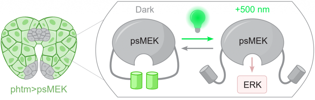
Optogenetics
System identification approaches are commonly used in engineering to infer simple yet predictive models of complex systems from their responses to time-dependent perturbations. Here, we apply this strategy at the whole organism scale, establishing a predictive model of commitment to metamorphosis in Drosophila. At this critical point in animal development, the larva stops feeding and proceeds to take on the adult form. The neuroendocrine circuits governing commitment to metamorphosis assess the growth and patterning programs, eventually triggering the production of systemic hormones that terminate growth and initiate metamorphosis. Previous studies of these circuits relied on relatively static genetic perturbations and starvation experiments. Here, we take advantage of optogenetic approaches in Drosophila to flexibly perturb a key signaling node within the endocrine gland in otherwise undisturbed larvae. We used this approach to infer parameters in a compact mathematical model and demonstrate that it makes accurate predictions of larval commitment to metamorphosis. Our work paves the way for quantitative studies of other juvenile-to-adult transitions, including mammalian puberty, which relies on strikingly similar mechanisms.
Cell lineage tracking
A key technique in live imaging embryonic development is accurately tracking cell lineages through multiple round of divisions…
<Schematic?>
<Pole cell tree video>
Mathematical modeling
Availability of quantitative data allows us to formulate and fit mathematical models of dynamical biological systems…
<Equations?>
<Embryo streaming video>